
Physical versus Biological Dosimetry
Comparison of electron spin resonance (ESR) estimation of tooth enamel dose and cytogenetic data of Hiroshima survivors
by Nori Nakamura, Chief, Department of Genetics
This article was originally published in RERF Update 9(1):3, 1998.
Using electron spin resonance (ESR) of tooth enamel to estimate atomic-bomb (A-bomb) gamma-ray dose received, we have recently examined 100 teeth donated from Hiroshima A-bomb survivors (Nakamura et al. 1998). To evaluate possible contamination from dental x-ray exposures, which affect mostly buccal surfaces, each tooth was divided into its buccal and lingual parts for subsequent independent enamel isolation and ESR measurement. Nearly 20 teeth showed considerably larger ESR-estimated doses in their buccal parts than in their lingual parts, which first seemed to indicate a considerably large contribution from dental x-ray exposure. However, it was found that most of these discrepant cases were incisors and canines. Because it is difficult to imagine that dental x-ray exposures affected mainly front teeth only and ultraviolet light exposure is reported to induce an ESR signal, the results are most likely attributable to solar light exposures (Romanyukha et al. 1996).
Next, cytogenetic examination was conducted for 61 of 69 tooth donors, and the results were compared with ESR estimates. Translocation frequency in lymphocytes and ESR-estimated dose for buccal and lingual parts of molars and lingual parts of front teeth, i.e., incisors and canines, showed a dose response for translocations as we would expect from in vitro gamma-ray irradiation experiments (Figure 1). However, the buccal parts of front teeth yielded definitely fewer translocations, and this is attributable to tooth-dose overestimation from solar light exposure contamination (Figure 2).
When the average doses in buccal parts were compared with those of lingual parts of the same teeth, the difference was largest for first incisors and declined following tooth position deeper in the mouth, i.e., 0.5 Gy for first incisors, 0.3 Gy for second incisors, 0.2 Gy for canines and small molars, and 0.04 Gy for large molars, including wisdom teeth. Because lingual parts of front teeth can also be affected by solar light exposures, although to a lesser extent than their buccal parts, these excess doses in buccal parts are probably minimal estimates attributable to solar light exposures.
We conclude that both tooth enamel ESR and cytogenetic tests of blood lymphocytes are useful measures for retrospective dose estimation of individuals. To be prudent, we should either avoid measuring front teeth or make an additional effort to eliminate solar light-induced ESR signals in using front teeth.
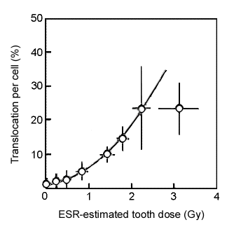
Figure 1. Translocation frequency versus ESR dose of lingual parts of molars, which are least likely affected by factors other than A-bomb gamma rays. Each point represents an average of five individuals, and bars represent standard deviations of the means. The line represents expected dose response, which is 70% of translocation yield measured by FISH (Lucas et al. 1995) because conventional Giemsa staining used in the present study is known to overlook 30% of translocations (Ohtaki et al. 1982).
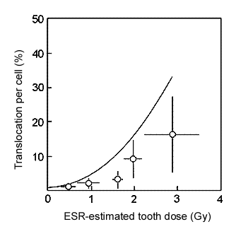
Figure 2. Translocation frequency versus ESR dose of buccal parts of front teeth, which are most likely affected by solar light exposures.
References about this subject