
Assessing Radiation Dose Recorded in Tooth Enamel
Electron-spin resonance measurements of extracted teeth donated by atomic-bomb survivors correlate fairly well with the lymphocyte chromosome-aberration frequencies for these same donors.
by N Nakamura,1 M Iwasaki,2 C Miyazawa,3 M Akiyama,4 AA Awa1
Departments of 1Genetics and 4Radiobiology, RERF; Departments of 2Dental Radiology and 3Preventive Dentistry, School of Dentistry, Ohu University, Kooriyama.
This article was originally published in RERF Update 6(2):6-7, 1994.
Radiation’s “fingerprints” remain in certain materials for quite a long time. For example, thermoluminescence has been used to measure atomic-bomb (A-bomb) gamma-ray doses using ceramic roof tiles in Hiroshima and Nagasaki more than 40 years after the bombings.
In the technique known as electron spin resonance (ESR) (also called electron paramagnetic resonance), radiation-induced radicals (ie, unpaired electrons) absorb microwaves when subjected to certain strengths of magnetic field. Different radicals (either atoms or molecules) have their own magnetic-field strength for the absorption. In liquid, radicals disappear quickly; but in solids, eg, in the calcified tissues of the human body such as bones and teeth, radicals can not freely move around and are much more stable. Because bones are continuously remodeled and are not readily accessible, mainly teeth have been used in ESR studies to date. (However, ESR dose estimates obtained from the bones of legs amputated due to radiation necrosis after accidental exposure have been reported [Desrosiers, Health Phys 61:859-61, 1991]). Enamel, the covering of the tooth surface, consists mostly of hydroxyapatite (a compound of crystalline structure consisting of calcium and phosphate) and is free from any metabolism. Tooth enamel is a unique inorganic body structure.
Although laboratory studies about ESR have been published since the 1960s, in the past decade ESR has received renewed attention. In Japan, M Ikeya of Osaka University has actively promoted ESR studies (eg, Ikeya et al, Jpn J Appl Phys 23:697-9, 1985; Ishii and Ikeya, Jpn J Appl Phys 29:871-5, 1990). A series of ESR studies on tooth enamel from mainly Nagasaki A-bomb survivors has been published by S Okajima’s research group at Nagasaki University (Tatsumi et al, J Hiroshima Med Assn 41:382-5, 1988 [in Japanese]; J Radiat Res 29:88, 1988 [English abstract]). These results have not been compared with individual Dosimetry System 1986 (DS86) dose estimates.
Current knowledge about ESR of tooth enamel
- It seems that CO33- radicals are being measured.
- Photon-energy dependence is evident, eg, 40-keV x rays generate an ESR signal per unit dose more than five times greater than cobalt-60 gamma rays (Tatsumi et al, J Hiroshima Med Assn 39:418-22, 1986 [in Japanese]; Iwasaki et al, Radioisotopes 40:421-4, 1991).
- Compared to gamma rays, neutrons are much less effective in producing ESR signals (Tatsumi, Filmbadge News 125:1-9, 1986 [in Japanese]; Iwasaki et al, unpublished observation, 1991).
- Observed ESR signal intensity is linearly proportional to the mass of enamel examined (Iwasaki et al, Ohu University Dental J 17:95-100, 1990).
- No dose-rate effect was observed after in vitro exposure to gamma rays with dose rates ranging from 225-0.33 R/min (Iwasaki et al, Radioisotopes 41:642-4, 1992).
- Irradiation of enamel samples in vitro, either in dry conditions or in water, produced identical ESR signal intensities (ibid).
- Enamel grain sizes of 0.5-1.4 mm in diameter are preferred (Iwasaki et al, Radioisotopes 42:470-3, 1993).
The Hiroshima ESR project
In Hiroshima, we began to request donations of extracted teeth from RERF’s Adult Health Study participants in 1986. Since that time, 300 teeth have been collected, but only about 30% were found to be suitable for enamel separation and subsequent ESR measurement.
We have learned that enamel must be carefully separated from dentin because dentin produces a large background signal and is very ineffective in producing a radiation-related signal. A disc-shaped diamond cutter with running water is currently used to isolate enamel after slicing a tooth. Imagine slicing the crust off of a piece of toast. This method, which is tedious and requires great skill, produces quite satisfactory results. (This method will be described in detail in the near future.)
One unsolved problem is how to evaluate the contribution to dose from dental x rays (which have an effective energy of 30 keV or less). As mentioned above, such low-energy photons are much more effective than cobalt-60 gamma rays and thus may contribute significantly to the ESR signal while actually contributing little to the dose. To assess dental x-ray dose, each tooth is divided in half–one half from the inside of the mouth and the other from the outside. Because panoramic photographs were not common in the past, we assume that most of the diagnostic dental x rays struck the outside of the tooth. Whenever a larger ESR signal is seen in the outer half than in the inner half of the tooth, we are particularly cautious in interpreting the results for that donor.
Preliminary results for 11 samples from 10 donors are presented in the Figure. Note that the current ESR measurements do not include any in vitro gamma irradiation of known doses. Thus, the ESR results may not necessarily correspond linearly with dose because each tooth may respond to radiation somewhat differently. Nevertheless, several interesting features are apparent. First, the two black circles connected with a line represent the results of two teeth from the same donor. The discrepancy seems mostly derived from dental x rays, because inner halves of the two teeth gave almost identical signal intensities (not shown) and are close to the lower value in the figure. Second, the chromosome-aberration data correlate quite well with the ESR data. In other words, no outliers are apparent. Third, one exceptional case has been noted. The tooth of a survivor whose DS86 assigned dose is 1.3 Gy and who has a 35% frequency of lymphocytes with chromosome aberration(s) showed no sign of radiation exposure! That particular sample was a wisdom tooth, known to be formed much later than other human teeth, and the tooth donor was 15 years of age at the time of the bombing. Because individual variation in the development of wisdom teeth is quite wide, we do not know if the tooth in question was really underdeveloped at the time of exposure in 1945. We hope to examine another type of tooth from this donor in the future.
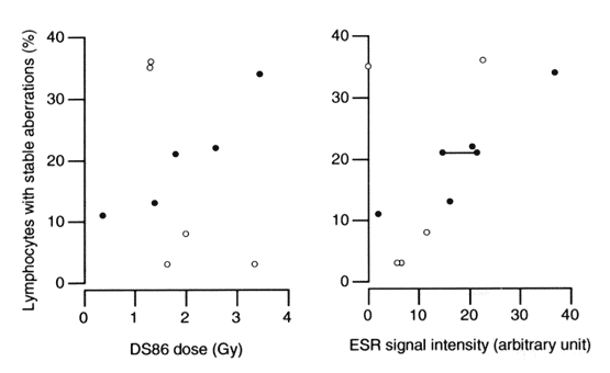
Figure. Left: frequency of lymphocytes bearing stable-type chromosome aberration(s) plotted against Dosimetry System 1986(DS86) total dose. Right: frequency of lymphocytes bearing stable-type chromosome aberration(s) plotted against ESR signal intensity of tooth enamel. Because each sample contains a different amount of enamel, the measured signal intensity was corrected by dividing with the weight. The black circles represent five cases that are close to the average in chromosome-aberration-dose response in our large cohort. The two black circles connected with a line represent the results of two teeth from the same donor. The five open circles are so-called outliers, although such individuals are not common. The ESR signal shown here is for outer halves of the samples.
Future prospects
In view of the applicability of ESR for dating fossil teeth, eg, from early Homo sapiens (Tiemei et al, Nature 368:55-6, 1994), it seems likely that tooth enamel can properly accumulate radiation doses imparted at extremely low-dose rates. This means that human teeth may be distinctive natural biodosimeters not only for acute radiation exposures but also for repeated small doses or chronic gamma-ray exposures of radiation workers and people residing in contaminated environments. At RERF, we intend to coordinate the ESR of tooth enamel with our lymphocyte chromosome-aberration studies that employ the fluorescence-in-situ-hybridization (FISH) method.