
Growth and Epilation of Human Hair in the Severe Combined Immunodeficient Mouse
Using an in vivo experimental model for human radiation exposure offers opportunities to understand the processes of radiation-induced pathogenesis.
by Seishi Kyoizumi, Department of Radiobiology, RERF
This article was originally published in RERF Update 7(2):4-5, 1995.
In 1988, J. M. McCune and his colleagues at Stanford University succeeded in implanting functional human fetal hematolymphoid organs, including thymus and lymph node tissues, into the severe combined immunodeficient (SCID) mouse (McCune et al, Science241:1632-9, 1988). When I read that article, an idea came to me: SCID mice implanted with human tissues (referred to as SCID-hu mice) could be irradiated with any sources and doses of radiation and thus serve as a suitable in vivo experimental model for human radiation exposure.
Although acute and late radiation effects have been documented in the atomic-bomb (A-bomb) survivors and persons exposed in radiation accidents, such information is limited by imprecise dose estimates and difficulty in obtaining biological samples for directly analyzing the process of radiation-induced pathogenesis. As a result, it has been difficult to achieve meaningful risk estimates.
In 1989, with these issues in mind, I spent a 2-year sabbatical leave at SyStemix Inc in Palo Alto, Calif, where Dr McCune was working. There I devised a SCID-hu mouse model for the radiobiological study of human hematopoiesis (Kyoizumi et al, Blood 79:1704-1, 1992; Blood 81:1479 88, 1993; Radiat Res 137:76-83, 1994). After returning to RERF in 1992, my colleagues and I have continued to explore the application of SCID-hu mice to human radiation-risk analyses from various points of view. Here, I will introduce briefly a SCID-hu skin model for the study of radiation-induced human epilation.
The SCID-hu mouse model for human epilation
Although epilation has long been recognized as one of the acute symptoms of ionizing radiation exposure, little is known about its dose response and mechanisms. By engrafting human skin tissue into SCID mice, we have developed an experimental model for radiation-induced epilation in humans. Skin tissue obtained from the head of a fetus who had died of malformation was engrafted onto the backs of SCID mice. The hair fell off several months after engraftment, but grew again and continued to grow for more than 1 year. About 5 months after engraftment, the skin grafts were locally irradiated with various doses of X rays. In the second week after irradiation, epilation was observed in the heavily irradiated group (exposed to greater than or equal to 3 Gy). In the third week, no epilation was observed up to doses of 1 Gy, and the epilation rate (the proportion of fallen hairs per unit area) was found to steeply increase at 2-3 Gy. More than half of the hair fell off after irradiation with X rays at greater than or equal to 3 Gy, and only less than 10% of the hair survived at 6 Gy (Figure 1). This is similar to the dose response for severe epilation reported in a previous study based on interviews with A-bomb survivors (DO Stram and S Mizuno, Radiat Res 117:93-113, 1989), supporting the validity of this SCID-hu mouse model.
The human hairs picked from the SCID-hu mice in the third and fourth week after irradiation were observed under a microscope. The hairs that fell off in the third week after high-dose irradiation (greater than or equal to 3 Gy) became thinner towards the roots and looked like the tips of needles. The histology of these damaged hair follicles showed that the bulb-like structure of normal hair had disappeared almost completely, and only the epithelial-like structure had survived. In the fourth week, the hairs surviving various doses of X-irradiation were clearly thin in the middle part and recovered gradually towards the root. The minimum diameter at the thinnest point of the hairs decreased dose-dependently with X-irradiation up to 3 Gy, and the mean diameter of the surviving hairs at 3 Gy was about 20 micrometers. These results suggest that the diameter of the hair decreases with radiation exposure, and epilation occurs as the hair breaks when its diameter decreases to approximately 20 micrometers or less. On the other hand, some radioresistant hairs survived even a 6-Gy irradiation.
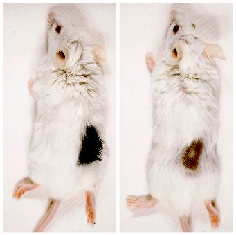
Figure 1. Left panel: a severe combined immunodeficient mouse implanted with human scalp tissue. Right panel: the human hair epilated 3 weeks after exposure to 6 Gy of X rays.
The diameter of these resistant hairs was about 25 micrometers and almost constant from the root to the tip. Histological analyses demonstrated that these resistant hairs were derived from hair follicles in the resting stage. Two to three months after epilation, hairs could regenerate even after a 6-Gy irradiation. However, the diameter of regenerated hairs was significantly thinner than that of nonirradiated hairs.
Implications for the mechanism of epilation
Figure 2 illustrates the hypothetical mechanism of radiation-induced human epilation. After irradiation, many proliferating cells in a hair matrix should be killed and decrease the activity of hair formation. In the high-dose range (greater than or equal to 3 Gy), the hair matrix is severely damaged or disappears, and the hair becomes thinner and finally breaks. In the low-dose range (less than or equal to 2 Gy), the hair also becomes thinner, but recovers its original thickness without hair breakage as the hair matrix recovers. Even in the case of high-dose irradiation, some follicle stem cells would survive in epithelial tissue derived from damaged follicles. The presence of follicle stem cells in the follicle epithelium has been proposed recently by a French research group (A Rochat et al, Cell76:1063-73, 1994). After their proliferation and differentiation, these stem cells would be able to regenerate follicle structure and reproduce a hair.
Our SCID-hu mouse system makes it possible to study radiation effects on human tissue in vivo, which can contribute significantly to our understanding of the pathogenesis of radiation-induced effects.
Figure 2. The hypothetical mechanism of
radiation-induced human epilation.
After irradiation, many proliferating cells in a hair matrix should be killed and decrease the activity of hair formation. In the high-dose range (greater than or equal to 3 Gy), the hair matrix is severely damaged or disappears, and the hair becomes thinner and finally breaks. In the low-dose range (less than or equal to 2 Gy), the hair also becomes thinner, but recovers its original thickness without hair breakage as the hair matrix recovers.