
Radiation-induced Mental Retardation: An Update
The authors discuss the abnormalities in brain architecture that may follow prenatal exposure to ionizing radiation.
by William J Schull, RERF Permanent Director, and Masanori Otake, RERF Department of Statistics
This article was originally published in RERF Update 3(4):3-4, 1991. The subject discussed in this article is more fully addressed in RERF TR 13-91.
Forty years have elapsed since it became apparent that prenatal exposure to the atomic bombings of Hiroshima and Nagasaki increased the frequency of mental retardation and the occurrence of an “atypically” small head. Much has since been learned about the stages in embryonic and fetal development at which these events are most likely to occur, and other subtle impairments of brain development have been identified, but the biologic picture remains incomplete. We are still uncertain about the “true” dose-response relationship, and whether a threshold does or does not exist. Neither of these uncertainties, despite their importance to regulatory agencies, seems likely to yield to epidemiological studies alone. If answers are to be found, they must lie in a better understanding of the cellular and molecular events involved in the development of the cortex. This will, in turn, require a closer integration of epidemiologic and experimental studies, particularly those of a molecular biological nature, than has yet been achieved.
A priori, the damage that has been seen could be attributable to a variety of biologic events working singly or in concert, and ranging from neuronal cell killing to impaired cellular reproduction, to mismanaged neuronal migration, to failures in normal synaptogenesis. But the relative importance of these different possible contributors remains unknown. Thus far, the most informative insights have come either from a small number of autopsy examinations or from the use of magnetic resonance imaging, a recently introduced noninvasive means of visualizing the living brain. Even here the information is quite limited, and can be summarized simply.
Autopsy findings
Four deceased survivors, who were prenatally exposed, have been autopsied. Two were mentally retarded; two were not. Only one received a high dose of more than 1 Gy. The others received doses less than 0.01 Gy. In the two with normal intelligence, the brains were of normal weight and the architecture appeared normal on visual inspection and microscopically. Both of the mentally retarded individuals, however, had brains that weighed substantially below normal. One had a brain weighing 840 g and the other 1,000 g (normal weight is about 1,450 g). Multiple slices through the larger brain, that of a female exposed in the 31st postovulatory week, revealed the usual pattern of gray and white matter and no evidence of swelling from the accumulation of fluid in the spaces between the brain cells which could have increased brain weight. She died at age 20 of heart failure.
The other mentally retarded individual, a male with the smaller brain, died at age 16, of acute meningitis of probable viral origin. If he had been carried to the normal termination of a pregnancy, he would have been exposed in the 12th postovulatory week, but given his birth weight (1,950 g), he was undoubtedly premature. His weight suggests that he was actually exposed at about the 8th or 9th week following fertilization since a full-term Japanese infant would now weigh about 3,200 g (but possibly somewhat less at the time of his birth in 1946). The estimated dose to his mother’s uterus was approximately 1.2 Gy.
Sections across the cerebrum revealed massive amounts of gray matter around the lateral ventricles, especially in the vicinity of the caudate nucleus, but also around the hippocampus. Microscopic examination of these misplaced gray areas revealed an abortive laminar arrangement of nerve cells, imitating the usual arrangement of the cortical neurons. The cerebellum and the curved ridges on the floor of the inferior horn of the lateral ventricle, the hippocampi, were normal visually and on microscopic study. However, the two protuberances on the under surface of the brain beneath the corpus callosum, called the mamillary bodies, were absent. These structures are presumed to play a role in memory since mamillary lesions are frequently associated with amnesia.
Misplaced gray matter was not observed in any of the other 3 autopsied cases, including the second mentally retarded individual. However, similar migratory errors have been seen in experimental rodents exposed to ionizing radiation at the equivalent time in development, and have been reported in other human fetuses exposed to massive amounts of ionizing radiation equally early in development.
Magnetic resonance imaging
Although the number of individuals that have been studied using magnetic resonance imaging is also small, several different anomalies of development have been seen. These correlate well with what is known of the embryological events transpiring at the time of their exposure. The findings on the neuroimaging of the 2 survivors exposed in the 8th or 9th week following fertilization do not differ qualitatively and are similar to those seen at autopsy in the case described above. When brain size is taken into account in both instances, there appears to be some increase in the size of the ventricles, but more importantly, there has been a failure of a significant number of neurons to migrate from the proliferative zone to their proper functional sites. One of these 2 individuals also exhibits an underdeveloped area in the left temporal region (see Figure 1). However, in these 2 cases, unlike the boy who was autopsied, the mamillary bodies are present and appear of normal size.
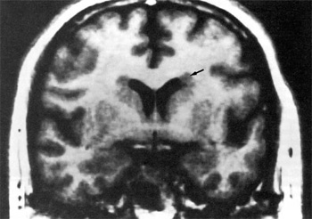
Figure 1. A coronal section through the brain of a survivor exposed prenatally in the 8th to 9th week following ovulation. Note the ectopias at the lateral margins of the ventricles above the caudate nucleus (arrow).
Ectopic gray matter has been seen in other instances of mental retardation not related to exposure to ionizing radiation, but its prevalence among mentally retarded individuals is not reliably known. The limited data that are available suggest that the nature of the migratory error could be different in the 2 instances. In the cases we describe, the failure occurs on both sides of the developing brain; whereas in nonradiation-related mental retardation it commonly involves only 1 side–although bilateral cases are known–and the ectopic area is often just beneath the cortex rather than around the ventricles. Ectopic gray matter is not invariably associated with mental retardation. Neuroimaging of individuals with the inherited Fragile X syndrome, among whom varying degrees of mental retardation commonly occur, has not revealed this defect. Among some 27 individuals who have been studied, just 8 were found to be abnormal. Seven of these individuals exhibited only a mild enlargement of the ventricles, but in 1 case a moderate, generalized dilation was seen. Autopsy studies have, however, disclosed abnormalities in dendritic spine morphology–very thin, long tortuous spines with prominent heads and irregular dilatations were noted. This suggests a developmental error occurring after migration was completed. Neuroimaging of 2 individuals exposed in the 12th to 13th postovulatory week reveals no conspicuous areas of misplaced neurons, but does show a faulty brain architecture. Again the abnormalities seen are surprisingly similar in the 2 cases. The prominently rounded elevations of the brain, the gyri, are enlarged, and the sulci, the furrows or trenches separating the gyri, are shallower than normal. One of the cases studied at this time exhibited a corpus callosum (the network of nerves which provides communication between the 2 halves of the brain) that was markedly smaller than normal (see Figure 2), and a poorly developed cingulate gyrus (the prominence lying immediately above the corpus callosum), suggesting an aberration in the development of the band of association fibers that passes over the corpus callosum. In both of these instances, the cistern involved in the recirculation of cerebrospinal fluid lying immediately behind and between the 2 lobes of the cerebellum, known as the cisterna magna, was markedly enlarged.
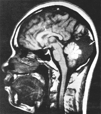
Figure 2. A sagittal section through the brain of a survivor exposed prenatally in the 12th to 13th week following ovulation. Note the enlarged cisterna magna (right arrow) and the deformed corpus callosum (left arrow).
Still later in development, at the 15th week, neither ectopic gray areas nor conspicuous changes in brain architecture were seen. Presumably the functional impairment that exists must be related to the connections that occur between neurons, possibly similar to the abnormality in the Fragile X syndrome described above. Experimental evidence shows that exposure at this time in the development of the brain in other primates leads to a diminished number of connections between neuronal cells. If all these connections have functional significance, then the diminution must compromise performance in some manner.
Although the observations just described are more informative than the simple determination of the frequency of mental retardation as a function of dose, they do not tell us what cellular or molecular events are impaired. However, when coupled with recent experimental findings, they are provocative. It is now clear that each cortical neuron has not only a designated date of birth, but a definite functional address. Since neuronal cells arise largely in specific proliferative, circumventricular zones, proper function implies migration. The process by which immature neuronal cells move from their sites of birth to those of their normal function is an active, timed phenomenon dependent on an interaction between their shapes, their surface membranes, and their guidance cells. Any damage to these membranes, however transitory, could conceivably impair migration. While there is presently no direct evidence of the effects of low doses of irradiation on the membranal properties of either neurons or the radial glial cells which guide them, experiments are underway which should provide some of the missing information. Finally, in interpreting the neuroimages that do not show evidence of ectopic gray matter, it warrants bearing in mind that the establishment of connections is essential to the survival of a neuron; those that do not form such connections die. The process of formation is competitive and neuronal cells that arrive late are obviously at a disadvantage.