
Radiation Cataract (Lens Opacity)
Radiation cataract causes partial opacity or cloudiness in the crystalline lens and results from damaged cells covering the posterior surface of the lens. Symptoms can appear as early as one or two years following high-dose exposure and many years after exposure to lower doses. It is unclear how frequently radiation cataracts advance to severe visual impairment, although we have documented in a recent study about a 20-30% excess at 1 Gy of cataracts that prompted cataract surgery. A low-dose threshold may exist below which radiation cataract does not arise, although our recent analyses suggest that there may not be a threshold, or if one exists, it is somewhere in the range of 0 to 0.8 Gy. The excess cataracts seen are of the types generally associated with radiation: posterior subcapsular and cortical cataracts. Figure 1 shows the relation between radiation dose and cortical opacity of lens.
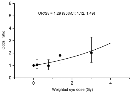
Figure 1. Cortical opacity of lens and radiation dose
Figure 2 describes how lens opacity is caused by radiation. There is a transparent layer of epithelial cells on the interior frontal side of the capsule that covers the lens. This layer maintains the function of the lens by slowly growing toward the center, achieved through cell division at the periphery (called the equator) of the lens. Because radiation is especially harmful to dividing cells, exposed cells at the equator are most prone to damage. For unknown reasons, damaged cells move toward the rear of the lens before converging on the center. Such cells prevent light from traveling straight forward, resulting in opacity.
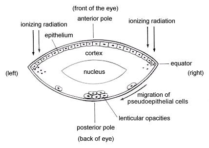
Figure 2. Lens opacity at the posterior subcapsular region caused by radiation
(from Effects of A-bomb Radiation on the Human Body, ed by HICARE in 1991,
courtesy of Bunkodo Co., Ltd., Tokyo)